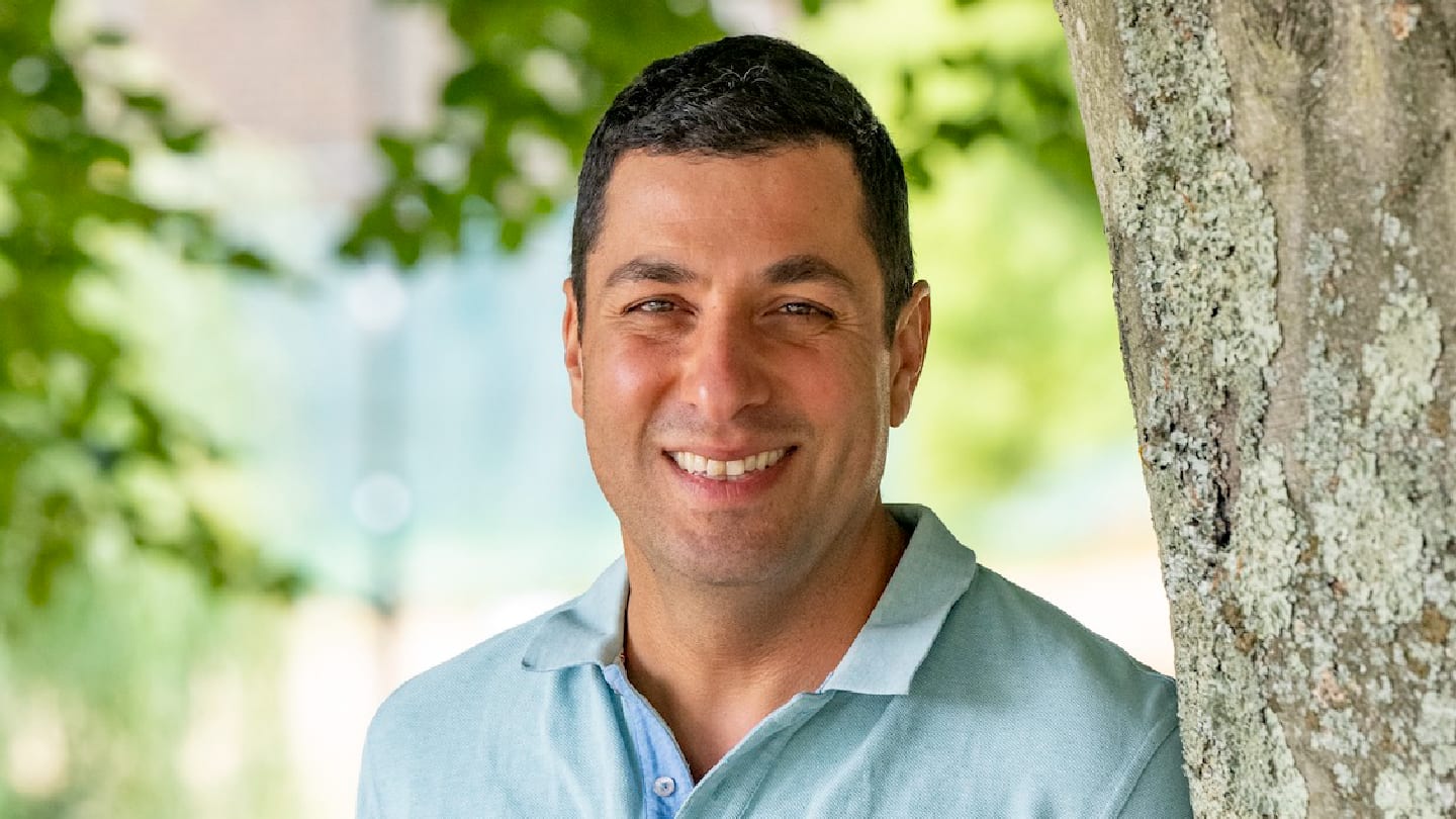
After making his Power List debut earlier this year, Ali Tavassoli’s work on the high-throughput intracellular production and screening of cyclic peptide libraries has been recognized with the Royal Society of Chemistry’s Interdisciplinary Prize.
Tavassoli’s work on protein–protein interactions can potentially address one of the most challenging areas in drug discovery: targeting the complex surfaces where proteins bind to each other to regulate vital biological functions. Such interactions are involved in many diseases, including cancer and viral infections, but they have traditionally been considered challenging because small molecules often cannot effectively disrupt them.
Tavassoli has developed an innovative approach, using cyclic peptides generated inside cells using genetically encoded libraries, that can block protein-protein interactions with high specificity. This opens the door to targeting previously inaccessible disease mechanisms and could lead to entirely new classes of therapeutics. His work not only expands the pool of viable drug targets but also provides powerful tools for discovering novel compounds faster and more precisely.
Here, we speak with Tavassoli to find out more about his research.
Congratulations on your recent prize-winning ventures! How does it feel?
It feels good! But for me, this isn’t just an award to myself, it’s an award for everyone who has contributed over the years to our shared scientific vision. We’ve been working to show that it's possible to create libraries of compounds inside cells and carry out high-throughput screening and drug discovery in a disease-relevant environment, rather than traditional approaches that screen in a biochemical buffer with recombinant proteins.
When we started this back in 2003 or 2004, it really was a bit of a crazy idea! The technology just hadn’t caught up with what we wanted to do. So, we began by engineering human proteins into bacteria and linking the survival of the bacterium to a human protein-protein interaction, then screening our compound libraries that way.
I feel that none of the awards I’ve received are mine alone. Anyone who stands up to claim sole credit for what is clearly a team effort hasn’t got it right. From the postdocs I worked with back when I was a postdoc, to those who trained me. This has always been a collaborative effort. Every student, every postdoc, every investor who believed in the vision has all played a part. The team at Curve Therapeutics now – our scientists, senior leaders, and investors – are all fantastic people who’ve chosen to come on this journey with us and dedicate themselves to this mission. The recognition belongs to all of them.
How has your approach advanced the identification of protein–protein interaction inhibitors?
Protein-protein interactions are thought to be difficult to inhibit. In some cases, there is a clear interaction between an alpha helix from one protein and a pocket in another protein, and in those cases, you can find inhibitors because there is a clear chemical motif that will point into the pocket from one protein to another.
But the majority of protein-protein interactions do not have a clear and obvious hotspot and are thought to be flat and featureless. Finding small molecules against these is considered to be very challenging.
In conventional approaches, researchers often produce the protein target in bacteria using a recombinant system. If the target is a human protein, it will lack the correct post-translational modifications. The protein is then placed into a biochemical buffer, which typically renders it static. If it requires a chaperone or another factor to adopt its active conformation, that won’t be present either.
Next, a screening method is applied – usually by washing a library over the protein. This could be a DNA-encoded library, phage display, or mRNA display. Compounds that bind are retained while others are washed away. After several rounds of washing, the remaining binders are identified by sequencing the DNA barcode or analyzing results from a multi-well plate assay. However, these assays often still use static protein in non-native conditions.
Our technique is fundamentally different. We use genetically encoded cyclic peptides that are cyclized head-to-tail through a process called intein splicing. The technique is called SICLOPPS and it enables us to generate libraries of hundreds of millions of cyclic peptides inside living cells. We also differ in the assays we use; rather than selecting hits based on binding affinity, we employ functional assays.
We express these libraries in disease-relevant mammalian cell lines, with each cell expressing one member of the library. We then engineer the system so that inhibition of a target – whether it's a transcription factor, a protein-protein interaction, autophagy, or any other process – produces a readable cellular phenotype. This lets us directly identify which cyclic peptides perturb the function of interest.
We separate the cells showing the desired phenotype and sequence the DNA in those cells to find the identity of the cyclic peptide that caused the desired effect. That peptide can be synthesized and tested independently, making our approach very different from traditional drug discovery.
Our cyclic peptides are just six amino acids in length, compared to the 12-20 amino acids typically found in mRNA or phage display peptides, which are already in "mini-protein" territory. This size difference matters because it’s one reason cyclic peptides have struggled to make it to market. Despite decades of development, cyclic peptide discovered by these methods have not successfully reached the market. One that did was withdrawn a few years ago.
Our smaller ring peptides are more akin to traditional small molecules. This lets us apply the same design principles used for small molecules. For instance, our hexameric peptides bind their targets through a continuous di- or tri-peptide motif – a “pharmacophore.” Thanks to the rigidity of the hexamer, we can model this pharmacophore effectively.
We can then “scaffold hop” by identifying small molecule backbones that mimic the cyclic peptide’s pharmacophore conformation. This enables us to move from a peptide scaffold to a small molecule scaffold, bringing us closer to clinical viability.
As both a professor at the University of Southampton, UK, and the CSO of Curve Therapeutics, how do you balance academic research with the goals of a biotech company?
I really enjoy both roles. The academic side gave me the freedom to explore ideas that would be considered too crazy in industry because of risk and timeline constraints. Biotech tolerates risk, but long timelines not so much.
Academia is often the source of innovation for biotech and pharma, which then develop it into real-world therapies. I became an independent academic in 2006, and some of the concepts we developed simply couldn’t have originated in a corporate setting.
I started Curve six years ago, but I’ve kept a reduced academic group and still teach one undergraduate course. My main focus now is Curve. I didn’t go into academia to do research for its own sake. I always wanted to apply what I did, and academia gave me the freedom to try the crazy stuff.
When I became a professor in 2014, I asked myself, “Is this it?” That’s when I started thinking about focusing my efforts on translating my research into the world. Biotech is fast-paced and dynamic. You can get things done quickly. If I’d stayed in academia, it would’ve taken me my entire career to get where we are today with Curve, where in five or six years, we’ve made enormous strides.
Can you share insights into how your platform is being used in collaboration with Merck, Sharpe & Dohme (MSD)?
The platform is unique. Not just because it allows screening inside cells, but because we use genetically encoded libraries. That means we can rapidly screen a billion cells with no robots required. That’s a game-changer. And our modality is different. We're working with six-amino-acid cyclic peptides – Microcycles – that others can’t easily use due to technical and chemical limitations.
It’s not just MSD – anyone who is interested in drug discovery against intracellular targets can see that this is different, and it’s accessible. Even those without a specialist scientific background appreciate the difference between screening in a test tube with a static protein versus screening in a human cell that reflects real disease biology.
You co-authored a report on a dual HIF-1 and HIF-2 inhibitor using the SICLOPPS platform. What’s the significance of that discovery?
HIF is crucial in solid tumours. It’s the protein that tells your cells, “Hey, you’re not getting enough oxygen. We need to adapt and send signals for new blood vessels.” That’s true for fetuses and tumors alike. Tumors grow quickly and outstrip their blood supply, known as hypoxia. HIF activation helps them survive and grow in this microenvironment.
HIF is a transcription factor with two subunits: HIF-alpha and HIF-beta. HIF-beta is constitutively expressed and always there, waiting in the nucleus. HIF-alpha is made and destroyed constantly – every few minutes – using oxygen-dependent hydroxylation as the signal for its degradation. When oxygen levels drop, HIF-alpha degradation stops and the protein is chaperoned into the nucleus, where it partners with HIF-beta, and changes the transcription of hundreds of genes to adapt to the low oxygen microenvironment.
From the moment it was discovered, HIF was recognized as a key cancer target. In 2013, we published the first HIF-1-specific inhibitor. Around the same time, a group in Texas published a HIF-2-specific inhibitor, which Merck eventually brought to market as belzutifan.
But many tumors are driven by both HIF-1 and HIF-2 – or they’ll switch between them – so we aimed to develop a dual inhibitor. That way, we could fully shut down hypoxia response in tumors. Our dual HIF-1/HIF-2 inhibitor remains the only one that targets the HIF-alpha/HIF-beta PPI directly. It’s one of the programs Curve is progressing toward the clinic.
How has your experience with the Royal Society of Chemistry influenced your research?
I was President of the Chemistry-Biology Interface Division at the Royal Society of Chemistry (RSC). I’ve always been drawn to multidisciplinary approaches to research. My PhD was in synthetic organic chemistry, then I did a postdoc using synthesis to understand biological mechanisms. This is how I got hooked on big-picture problem solving.
If you work in just one discipline, your tools are limited. But when you span fields, you can ask: “What’s the best way to tackle this problem?”
I spent five years in Steve Benkovic’s lab at Penn State University, for most of that time re-training in biology. When I joined Southampton in 2006 as an independent academic, they were forward-thinking enough to support cell culture labs in a chemistry department, which was rare at the time, that allowed my lab to take a multidisciplinary approach to our projects from the very start.
That mindset of bringing together the right tools regardless of discipline has guided my research since. Through my roles at the RSC, I’ve worked to highlight interdisciplinary science to funders and government. It’s been personally enriching and, I hope, helpful to the community as well.
What future opportunities or challenges do you see in intracellular target discovery?
The academic group is still going strong! We’re still doing crazy stuff and we have a few exciting papers coming out soon.
One involves using cyclic peptides to discover new antibiotics. Another explores targeted protein degradation. And there’s a third project I can’t talk about just yet, but trust me, it’s exciting!
I would like to give credit to my students and postdocs who believed in these wild ideas and bring them to life. Without them, an idea remains just that. It is particularly satisfying that we’re continuing to push the boundaries of what we do. I feel incredibly lucky to be surrounded by such amazing people who help turn these “crazy” ideas into reality.


